10 Tips for Choosing a Medical Ultrasound Transducer
FLUID HANDLING EXPERTS

Blog, all our news

10 Tips for Choosing a Medical Ultrasound Transducer

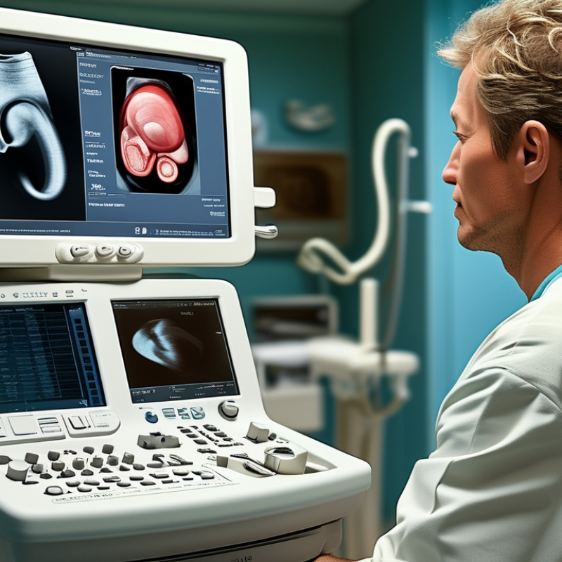
Choosing a Medical Ultrasound Transducer is a clinical decision, not merely a purchasing task. The probe affects image quality, examination speed, patient comfort, and diagnostic confidence. A cardiac transducer cannot replace a high-frequency linear probe for superficial vessels. Likewise, an abdominal probe may struggle with small tendons or shallow lesions.

Dr. K. Kirk Shung, a respected ultrasound technology researcher, described ultrasound as “noninvasive, safe, and relatively inexpensive.” That principle still matters. A dependable transducer should support safe scanning, accurate imaging, and consistent performance during demanding examinations. It should also match the ultrasound system, intended specialty, patient population, and daily workload. Specifications matter. So does handling.

This guide presents ten practical tips for choosing a Medical Ultrasound Transducer. It considers frequency range, probe design, bandwidth, footprint, ergonomics, compatibility, durability, cleaning requirements, warranty support, and total cost. Real clinical details guide the discussion, from scanning between narrow ribs to examining a superficial tendon under firm contact. However, no checklist removes every uncertainty. A probe that performs well in one clinic may feel poorly balanced in another. User training, maintenance habits, and machine settings can change the result.

That is easy to overlook.

Experienced sonographers should test the transducer before purchase whenever possible. They can assess cable flexibility, grip comfort, image uniformity, and sensitivity using familiar cases. Careful selection does not guarantee perfect images. It improves the chances of obtaining reliable ones.
10 Tips for Choosing a Medical Ultrasound Transducer

Define Clinical Depth: Use 2–5 MHz for Deep and 5–15 MHz for Superficial Imaging

10 Tips for Choosing a Medical Ultrasound Transducer

Define Clinical Depth: Use 2–5 MHz for Deep and 5–15 MHz for Superficial Imaging

Transducer frequency should match the anatomy, not personal preference. For abdominal, pelvic, or obstetric imaging, 2–5 MHz usually provides deeper penetration. Higher frequencies lose energy faster in tissue. The commonly cited attenuation estimate is about 0.5 dB/cm/MHz in soft tissue, according to ultrasound physics guidance from the AIUM. A 5 MHz beam therefore attenuates faster than a 2 MHz beam.

For thyroid, breast, vascular, and musculoskeletal studies, 5–15 MHz often produces sharper superficial detail. The ACR–AIUM–SPR–SRU ultrasound practice parameters emphasize selecting equipment according to the examination and target depth. A superficial linear transducer can reveal tendon fibers clearly. It may fail, however, when the target lies beneath thick tissue. Depth matters more than attractive resolution.

I have seen users choose the highest frequency available. That decision sometimes creates a clean image with missing anatomy. Lower frequency probes can look less detailed, yet they may show the complete lesion. Measure the expected depth before scanning. Leave practical penetration margin. Patient habitus, edema, and probe pressure can change the result. Technical reports often simplify frequency ranges, but real examinations are rarely so tidy. A dual-frequency option may help when anatomy varies during the same session. Still, operators should confirm image quality, focal position, and penetration on the patient, not only on a specification sheet.

Select Probe Geometry: Compare Linear, Curved, Phased, and Endocavity Designs

Choosing a medical ultrasound transducer starts with anatomy, not habit. Probe geometry controls contact, field of view, penetration, and maneuverability. A linear transducer produces a rectangular image and works well for superficial vessels, tendons, thyroid tissue, and guided needle placement. Its high-frequency footprint shows crisp detail, but depth falls quickly in larger patients. Keep that limitation visible.

A curved transducer offers a wider field at depth through a smaller contact edge. It suits abdominal, obstetric, and general pelvic examinations, especially when tissue lies beneath a broad surface. However, the curved image can make needle angles and lateral measurements less intuitive. A phased-array design uses a small footprint and sector-shaped beam. It can fit between ribs and examine the heart, but the image narrows near the skin and demands careful probe positioning. Small adjustments matter.

Endocavity transducers support transvaginal or transrectal examinations, where close contact provides detailed images of nearby structures. Their geometry improves access, yet comfort, cleaning workflow, protective covers, and documented infection-control procedures must guide selection. Compare frequency range, footprint size, steering controls, cable flexibility, and system compatibility. Ask clinicians to test each probe on typical patients, not only a training phantom. That exposes awkward grips and missed anatomy. I once favored a compact footprint too quickly; it handled tight spaces well but felt tiring during longer studies. Real workflow can challenge a technically impressive specification.

Evaluate Doppler Performance: Check PRF, Bandwidth, Sensitivity, and Frame Rate

Tip 1: Evaluate PRF in real scanning conditions. A higher PRF can reduce aliasing when blood moves quickly. However, it may limit detection of slower flow. Check whether the transducer supports practical adjustments for different vessels and depths. Do not trust the maximum number alone.

Tip 2: Examine bandwidth and sensitivity together. Broad bandwidth supports flexible imaging and Doppler applications. Sensitivity matters when signals are weak, especially in deep tissue or low-flow studies. Ask for phantom data, then compare it with supervised clinical scans. A specification sheet is useful, but it is not the whole story. Small differences may become obvious near a vessel wall.

Tip 3: Check frame rate while Doppler is active. Color Doppler can become sluggish when the region of interest is wide or the imaging depth increases. A responsive frame rate helps capture changing flow and improves timing during examinations. Test several box sizes, depths, and pulse repetition settings. I have seen excellent sensitivity paired with disappointing motion response. That trade-off deserves honest discussion.

A reliable evaluation should include experienced sonographers, repeat measurements, and documented conditions. Compare spectral waveforms, color fill, noise, and signal stability. Consider the intended examinations, not only laboratory performance. The best choice may not have the highest bandwidth or frame rate. It should deliver consistent Doppler information in the hands of your clinical team. Even careful testing can miss workflow problems, so allow time for review after daily use.

Verify Safety Compliance: Review FDA MI ≤1.9, TI ≤6.0, and IEC 60601-2-37

When choosing a medical ultrasound transducer, safety verification should begin with measurable output limits. The U.S. FDA’s diagnostic ultrasound guidance identifies a Mechanical Index (MI) limit of 1.9 and a Thermal Index (TI) limit of 6.0 for applicable diagnostic systems. These values are not decorative screen numbers. They reflect potential mechanical and thermal effects during scanning.

Check the transducer with its intended console, presets, and operating modes. Confirm that MI and TI remain visible and accurate during high-output imaging, Doppler use, and prolonged examinations. IEC 60601-2-37 specifies particular safety requirements for diagnostic ultrasound equipment, including output display and protective design. The AIUM’s ultrasound safety guidance also supports the ALARA principle: use the lowest exposure that produces a useful image.

A practical review should include test records, software version, calibration status, and probe condition. Look for cracked housings, damaged acoustic lenses, or unstable temperature readings. Small defects can create large uncertainties. I have found that teams sometimes check the console but overlook transducer-specific behavior. That is a weakness. Safety claims should be verified under realistic clinical presets, not only laboratory defaults. Ask for documented test results, risk controls, and evidence of compliance with FDA expectations and IEC requirements before purchasing.

10 Tips for Choosing a Medical Ultrasound Transducer - Verify Safety Compliance: Review FDA MI ≤1.9, TI ≤6.0, and IEC 60601-2-37
Tip Evaluation Dimension Verifiable Requirement or Reference Data Recommended Evidence Review Method
1 Clinical application Confirm that the transducer is intended for the required examination, such as abdominal, vascular, cardiac, obstetric, or superficial imaging. Intended-use statement, product specifications, and applicable regulatory labeling. Match the transducer's declared applications with the department's examination protocols.
2 Frequency range Use lower frequencies for deeper penetration and higher frequencies for superficial structures. A typical diagnostic range may be approximately 2–15 MHz, depending on transducer design. Published operating-frequency range and examination-depth data. Check whether the frequency range supports the target anatomy and required image depth.
3 Acoustic output safety Review displayed MI ≤ 1.9 and TI ≤ 6.0 against the applicable FDA-cleared labeling and system configuration. MI represents mechanical effects; TI estimates potential tissue heating. Acoustic-output table, system user manual, regulatory submission documents, and on-screen output display. Verify the highest applicable MI and TI values across supported operating modes, presets, and output-power settings.
4 IEC safety compliance Confirm evaluation against IEC 60601-2-37, which specifies particular requirements for the basic safety and essential performance of ultrasonic medical diagnostic and monitoring equipment. Current test report or certificate covering IEC 60601-2-37, together with the applicable IEC 60601-1 safety documentation. Check certificate scope, tested configuration, standard edition, and report validity.
5 Electrical protection Confirm appropriate protection against electric shock, leakage current, and applied-part hazards for the intended clinical environment. Electrical-safety test report, applied-part classification, and equipment protection rating. Verify that the transducer and compatible ultrasound system are tested as the intended medical equipment configuration.
6 Surface temperature Review probe-surface temperature limits and any special restrictions for endocavitary, neonatal, pediatric, or prolonged-contact examinations. Thermal test results, operating instructions, and cleaning or warm-up requirements. Confirm that maximum surface temperature and contact-time instructions are suitable for the intended patient population.
7 Ingress protection and durability Check the stated protection against fluid ingress, impact, cable bending, connector stress, and repeated clinical handling. Ingress-protection rating where applicable, environmental specifications, and durability test results. Compare the rated protection with operating conditions such as bedside use, fluid exposure, and transport frequency.
8 Disinfection compatibility Ensure that approved cleaning and disinfection methods are compatible with the transducer materials, cable, lens, and connector. Validated reprocessing instructions listing compatible disinfectants, concentration, contact time, and temperature. Confirm that the facility's infection-control procedure does not exceed the stated chemical or mechanical limits.
9 Image quality and performance Assess axial resolution, lateral resolution, penetration, uniformity, dead zone, frame rate, and sensitivity using a suitable test object. Acceptance-test results, phantom measurements, and representative clinical images. Compare measurements with the procurement specification and establish a baseline for periodic quality control.
10 Compatibility and lifecycle support Verify connector compatibility, supported imaging modes, software requirements, calibration support, warranty terms, and replacement availability. Compatibility matrix, service documentation, calibration procedure, warranty conditions, and maintenance schedule. Confirm that the complete transducer-system combination is authorized, serviceable, and supportable throughout the planned lifecycle.
Safety limits and labeling requirements can depend on the ultrasound system, operating mode, transducer type, intended use, and applicable regulatory pathway. Final acceptance should be based on the current device documentation, applicable FDA requirements, and the complete IEC test scope.

Assess Workflow Value: Confirm Compatibility, IPX7 Protection, Sterilization, and Cost

A medical ultrasound transducer should fit the real workflow, not just the equipment list. In ultrasound service work, I have seen a technically excellent probe slow examinations because its connector, presets, or imaging mode did not match the system. Confirm compatibility with the manufacturer’s current documentation and your department’s software version. Check frequency range, footprint, cable length, and intended examinations. A small mismatch can create daily delays.

IPX7 protection is useful when a transducer may face fluid exposure or careful immersion cleaning. However, IPX7 does not automatically make every probe suitable for full sterilization. Review the instructions for use, approved disinfectants, exposure times, and temperature limits. Ask whether the probe supports high-level disinfection or only low-level cleaning. Keep records.

Cleaning takes time.

Cost should include more than the purchase price. Compare expected service life, repair access, downtime, cleaning supplies, and replacement availability. A lower-priced transducer may become expensive if its cable fails early or its design complicates disinfection. I once focused too heavily on acquisition cost and underestimated staff training time. That was a poor assumption. Invite sonographers, infection-control staff, and biomedical engineers to test the workflow. Let them handle the probe with gloves, connect it repeatedly, and inspect its surfaces under ordinary room lighting. Their practical feedback may reveal weaknesses that specifications conceal. Verification against applicable clinical and electrical safety requirements remains essential.

10 Tips for Choosing a Medical Ultrasound Transducer — Assess Workflow Value

Typical operating-frequency ranges can help match a transducer to clinical workflow. Confirm system compatibility, IPX7 protection, approved sterilization methods, service requirements, and total cost before purchase.

Frequency ranges are typical clinical values: lower frequencies generally support deeper penetration, while higher frequencies provide greater superficial detail. Actual specifications vary by model and application.

Copyright 2025 Fluidra S.A. | Created by BCM Marketing Agencia Privacy Policy Legal note | Cookie policy |

Technical cookies are strictly necessary for our website to work and you can navigate through it. These types of cookies are those that, for example, allow us to identify you, give you access to certain restricted parts of the page if necessary, or remember different options or services already selected by you, such as your privacy preferences. Therefore, they are activated by default, your authorization not being necessary in this regard. Through the configuration of your browser, you can block or alert the presence of this type of cookies, although such blocking will affect the proper functioning of the different functionalities of our website.


The analysis cookies allow us to study the navigation of the users of our website in general (for example, which sections of the page are the most visited, which services are used most and if they work correctly, etc.). From the statistical information on navigation on our website, we can improve both the operation of the page itself and the different services it offers. Therefore, these cookies do not have an advertising purpose, but only serve to make our website work better, adapting to our users in general. By activating them you will contribute to this continuous improvement. You can enable or disable these cookies by checking the corresponding box, being disabled by default.


Functionality cookies allow us to remember your preferences, to personalize certain characteristics and general options of our website, every time you access it (for example, the language in which the information is presented to you, the sections marked as favorites, your browser type, etc.) Therefore, this type of cookies does not have an advertising purpose, but by activating them you will improve the functionality of the website (for example, adapting to your type of browser) and the customization of it based on your preferences (for example, by presenting the information in the language that you have chosen in previous occasions), which will contribute to the ease, usability and comfort of our page during your navigation. You can enable or disable these cookies by checking the corresponding box, being disabled by default.


Advertising cookies allow us to manage the advertising spaces included in our website based on criteria such as the content shown or the frequency in which the ads are displayed. For example, if you have been shown the same ad several times on our website, and you have not shown a personal interest by clicking on it, it will not appear again. In summary, by activating this type of cookies, the advertising shown on our website will be more useful and diverse, and less repetitive. You can enable or disable these cookies by checking the corresponding box, being disabled by default.


Behavioral advertising cookies allow us to obtain information based on the observation of your browsing habits and behaviors on the web, in order to show you advertising content that best suits your personal tastes and interests. To understand it in a very simple way, we will give you a fictitious example: if your last searches on the web were related to suspense literature, we would show you advertising about suspense books. Therefore, activating this type of cookies, the advertising that we show you on our website will not be generic, but will be oriented to your searches, tastes and interests, therefore adjusting exclusively to you. You can enable or disable these cookies by checking the corresponding box, being disabled by default.
To understand it in a very simple way, we will give you a fictitious example: if your last searches on the web were related to suspense literature, we would show you advertising about suspense books. Therefore, activating this type of cookies, the advertising that we show you on our website will not be generic, but will be oriented to your searches, tastes and interests, therefore adjusting exclusively to you. You can enable or disable these cookies by checking the corresponding box, being disabled by default.

SAVE

We use our own and third-party cookies to ensure the proper functioning of the website and to analyse our services, improve your experience on our website and show you advertisements related to your interests according to profiling based on your browsing habits (e.g. websites visited). You can accept all cookies by clicking "ACCEPT" or set or reject them by clicking here. If you would like to know more about the use of cookies, please see our COOKIE POLICY.


ACCEPT ALL
Reject no essentials